Sukanya Dej-adisai ![]() ,
Kedsaraporn Parndaeng,
Chatchai Wattanapiromsakul
,
Kedsaraporn Parndaeng,
Chatchai Wattanapiromsakul
For correspondence:- Sukanya Dej-adisai Email: sukanya.d@psu.ac.th Tel:+6674428220
Received: 15 March 2015 Accepted: 7 January 2016 Published: 31 March 2016
Citation: Dej-adisai S, Parndaeng K, Wattanapiromsakul C. Determination of phytochemical compounds, and tyrosinase inhibitory and antimicrobial activities of bioactive compounds from Streblus ilicifolius (S Vidal) Corner. Trop J Pharm Res 2016; 15(3):497-506 doi: 10.4314/tjpr.v15i3.10
© 2016 The authors.
This is an Open Access article that uses a funding model which does not charge readers or their institutions for access and distributed under the terms of the Creative Commons Attribution License (http://creativecommons.org/licenses/by/4.0) and the Budapest Open Access Initiative (http://www.budapestopenaccessinitiative.org/read), which permit unrestricted use, distribution, and reproduction in any medium, provided the original work is properly credited..
Purpose: To determine the phytochemical content, and tyrosinase inhibitory and antimicrobial activities of the wood from Streblus ilicifolius (S. Vidal) Corner
Methods: The dried wood of S. ilicifolius (8.70 kg) was extracted by maceration to give petroleum ether, ethyl acetate, ethanol and water extracts, respectively. Dopachrome method was used to determine antityrosinase activity. Agar disc diffusion and modified broth microdilution methods were used to determine antimicrobial activity. Chromatographic techniques were used for phytochemical investigation. The structures elucidation of isolated compounds were identified by physical properties and spectroscopic data including UV, IR, NMR and MS data and confirmed by comparison with previously reports.
Results: The ethanol extract exhibited tyrosinase inhibition and antimicrobial activity against the Gram-positive bacteria, Staphylococcus epidermidis and S. aureus. Phytochemical investigation showed five compounds, namely, (E)-2,4-dihydroxy-3-(3,7-dimethyl-2,6-octadienyl) benzaldehyde (1), p-hydroxybenzoic acid methyl ester (2), umbelliferone (3), moracin M (4), trans-resveratrol (5). Compound 4 exhibited tyrosinase inhibition with half maximal inhibitory concentration (IC50) of 67.69 µg/ml, while compound 1 displayed strong activity against S. epidermidis, S. aureus and methicillin-resistant S. aureus (MRSA) with minimum inhibitory concentration (MIC) of 8, 4 and 8 µg/ml, respectively and minimum bactericidal concentration (MBC) of 32, 16 and 64 µg/ml, respectively.
Conclusion: This is the first report of the biological activities and phytochemical composition of S. ilicifolius and the results indicate the high potentials of the plant for commercial applications such as in facial whitening and anti-acne cream.
Introduction
Melanin biosynthesis or melanogenesis initiated from L-tyrosine hydroxylated to L-dihydroxyphenylalanine (L-Dopa), then oxidation of L-Dopa to its corresponding o-dopaquinone, catalyzed by tyrosinase enzyme. o-Dopaquinone can be divided into two different types of reaction to produce eumelanin and pheomelanin [1,2]. The major function of melanin is to protect against ultraviolet (UV) radiation. However, excessive level of melanin or hyperpigmentation due to a darker or uneven of skin color, age spots, melasma and sites of actinic damage [1].
Tyrosinase inhibitors are becoming increasingly important contain in the treatment products of some dermatological disorders, associate with melanin hyperpigmentation and contain in cosmetic for skin whitening products [1]. Many problems from current whitening cosmetics such as ochronosis, allergic and irritant contact dermatitis, melanocyte toxicity and carcinogenicity [3] cause to find out the new natural whitening agents.
Pathogenic bacteria and dermatophyte fungus are a major cause of human skin disease. Moreover, some microorganisms can be stimulating the melanogenesis [4-7]. Antibiotics are the choice of treatment. However, the use of antibiotics may lead to drug resistance of many bacterial strains. Development of new antimicrobial compounds for resistant organisms is becoming critically important [8].
It is prompt search to find alternative agents from plants that have already been used topically to treat hyperpigmentation in skin of color [1,3,9]. The discovery of tyrosinase inhibition from medicinal plants is alternatives may provide leads for anti-pigmentation compounds and development to whitening and anti-browning agents.
In Thailand, there are many species of the plants in the genus Streblus, but only S. asper has been reported in traditional uses, and also their pharmacological activities and chemical constituents. However, in this study we have the focus on biological activities of S. ilicifolius wood extract. The extract showed the potential of antityrosinase and antimicrobial activities. The aim of this study was to evaluate some biological activities and chemical constitution of S. ilicifolius.
Methods
Plant material
140 samples from 79 Thai medicinal plant extracts of Rajjaprabha Dam, Surat Thani Province were screened for antityrosinase activity. S. ilicifolius (S. Vidal) Corner was collected from Rajjaprabha Dam, Surat Thani Province. The plant was identified by Ms. Nannapat Pattharahirantricin, botanist at Plant taxonomy Office of the Forest Herbarium, Department of National Parks, Wildlife and Plant Conservation, Bangkok, Thailand. The voucher specimen number of this plant was SKP 117 19 09 01. It was collected at the Department of Pharmacognosy and Pharmaceutical Botany, Faculty of Pharmaceutical Sciences, Prince of Songkla University, Songkhla, Thailand.
Extraction and isolation
The dried wood of S. ilicifolius (8.7 kg) was chopped, ground and macerated with petroleum ether three times (3 days, each). The filtrates were pooled and evaporated under reduced pressure at temperature not exceeding 40 oC. to yield a petroleum ether extract. Then the marc was extracted three times with ethyl acetate and ethanol (3 days, each). It follows an extraction with boiling water. Removal of the solvents gave an ethyl acetate, ethanol and H2O extract, respectively. The ethanol extract, showed the potential activities of antityrosinase and antimicrobial was selected for phytochemical investigation. The extract was fractionated by quick column chromatographic technique using a sintered glass-filter column of silica gel 60 (Merck). It was performed a gradient elution using mixtures of chloroform and ethyl acetate (10:0 to 2:8). Finally ethyl acetate was used to wash the column until the eluates were diluted and clear. The result was 13 fractions A-M. Repeated column chromatography of fraction A, gradient elution was performed using mixtures of hexane and ethyl acetate (9:1 to 1:1), given to 15 subfractions. The subfraction A-7 was purified by repeat column chromatography to obtain compound 1. Repeated column chromatography of fraction B, seperation was carried out with gradient elution, using mixtures of hexane and ethyl acetate (7:3 to 1:1), to give 8 subfractions. The subfraction B-2 was further fractionated by repeated column chromatography to obtain compound 2 and 3. Repeated column chromatography of fraction E, gradient elution was performed using mixtures of chloroform and methanol (10:0 to 1:1). 12 subfractions were isolated. The subfraction E-8 showed the major spot was further fractionated by repeated column chromatography and crystallized (chloroform-methanol, 9:1) to obtain compound 4 and 5.
General experimental procedures
The structure of compounds was characterized by NMR spectroscopy and mass spectrometry. 1H and 13C NMR, COSY, HMQC and HMBC spectra were recorded on Varian Unity Innova spectrometer operating at 500 MHz for 1H-NMR and 125 MHz for 13C-NMR and Bruker Avance III (1H-NMR 300 MHz and 13C-NMR 75 MHz). Mass spectra were measured on Thermo Finnigan MAT 95 XL mass spectrometer. The EI-MS of the compound was determined by GC-MS analysis using an Agilent 7890A GC system interfaced to an Agilent 5975C MSD operating at 70 eV, ion source temperature 230 °C, interface temperature 280 °C. A split injection (1 µl injection volume, split ratio, 20:1) at 260 °C injector temperature was utilized. A fused silica capillary column 5 % phenyl 95 % methyl polysiloxane (HP-5MS 30 m x 250 µm x 0.25 µm, Agilent J & W, USA) was used. The temperature program was as follows: 1 min at 60 °C, then to 220 °C at 10 °C/min, then to 300 °C at 4 °C/min, temperature kept at 300 °C for 10 min. The carrier gas was helium 5.6 at a flow rate 1.2 ml/min. Data acquisition was performed with Agilent GC/MSD ChemStation Version E.02.00 for the mass scan range 40-700 u. Column chromatography was carried out using silica gel 60 (particle sized 0.040-0.063 mm). All reaction steps were checked by thin-layer chromatography (TLC). The analyses were performed on pre-coated silica gel 60 F254 plates. Spots on TLC were visualized under UV absorption (254 and 366 nm). For this purpose, the plate was sprayed with anisaldehyde/H2SO4 reagent followed by heating.
Determination of antityrosinase activity
Dopachrome method was used to determine antityrosinase activity, using L-Dopa as the substrate [10]. Dopachrome is one of the intermediate substrate in melanogenesis, this method used the L-Dopa as a substrate. The red color of dopachrome from the oxidation of L-Dopa can be detected by visible light at 492 nm. Antityrosinase activity was performed in 96 well microplate. The samples were dissolved in DMSO to concentration of 200 µg/ml. The first 20 µL of the samples solution were mixed with phosphate buffer (pH 6.8) and 20 µL of mushroom tyrosinase solution (203.3 U/ml) and incubated at 25 oC for 10 min. Then 20 µL of L-Dopa (0.85 mM) was added. The visible absorption was measured at 492 nm. The solution was incubated at 25 oC for 20 min. After incubation the amount of dopachrome in the reaction was measured at 492 nm again. Tyrosinase inhibition (T) was calculated as in Equation 1.
T (%) = {[(A - B) - (C - D)]/(A - B)}100 …….. (1)
where A is the difference of optical density before and after incubation of control, B is the difference of optical density before and after incubation of blank control, C is the difference of optical density before and after incubation of test sample, and D is the difference of optical density before and after incubation of blank sample.
The concentration of the pure compound was measured which caused a 50 % inhibition of tyrosinase activity (IC50). Kojic acid and water extract of Artocarpus lakoocha wood were used as positive controls while DMSO was used as negative control.
Determination of antimicrobial activity
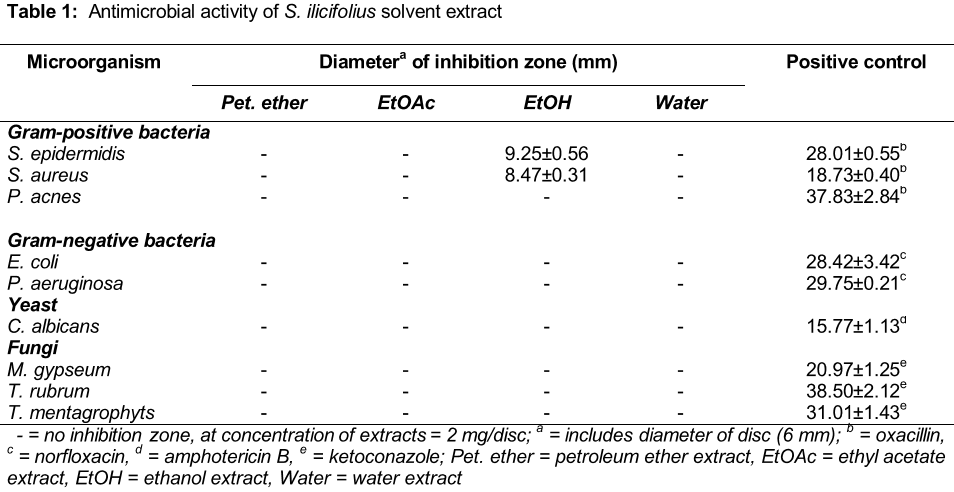
Microorganisms; Staphylococcus aureus (ATTC 25923), Staphylococcus epidermidis (TISTR 517), Propionibacterium acnes (DMST 14916) and Methicillin-resistant Staphylococcus aureus (MRSA 1350II 06) were alternate for gram positive bacteria. Escherichia coli ATCC35218 and Pseudomonas aeruginosa ATCC10145 were alternate for gram negative bacteria. Candida albicans (TISTR 5779) was alternate for yeast and Microsporum gypseum, Trichophyton rubrum and Trichophyton mentagrophytes were alternate for fungi. The preliminary screening of anti-microbial activity was used agar disc diffusion method [11]. The pure compounds were determined for minimum inhibitory concentration (MIC) and minimum bactericidal concentration (MBC) in 96 well microplate by modified broth microdilution method [12,13]. Oxacillin, norfloxacin, amphotericin B and ketoconazole were used as positive controls for gram positive bacteria (except MRSA using vancomycin), gram negative bacteria, yeast and fungi, respectively.
Results
Antityrosinase activity
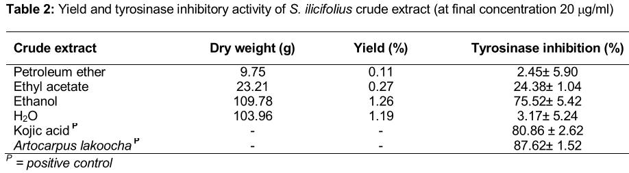
From 140 samples, S. ilicifolius ethanol wood extract showed the highest activity of antityrosinase with 69.05 ± 5.00 %.
Antimicrobial activity
The ethanol extract of the wood from S. ilicifolius showed the potential antibacterial effects against S. epidermidis and S. aureus with inhibition zone 9.25 ± 0.56 and 8.47 ± 0.31 mm, respectively ().
Extraction of S. ilicifolius wood
The dry weight, % yield (base on dried wood) and % tyrosinase inhibition of these crude extracts from the wood of S. ilicifolius are shown in . The ethanol extract was the most interesting fraction because of the potential effects of antityrosinase and antimicrobial activities. So it was selected for isolation of the chemical constituents.
Structure of isolated compounds
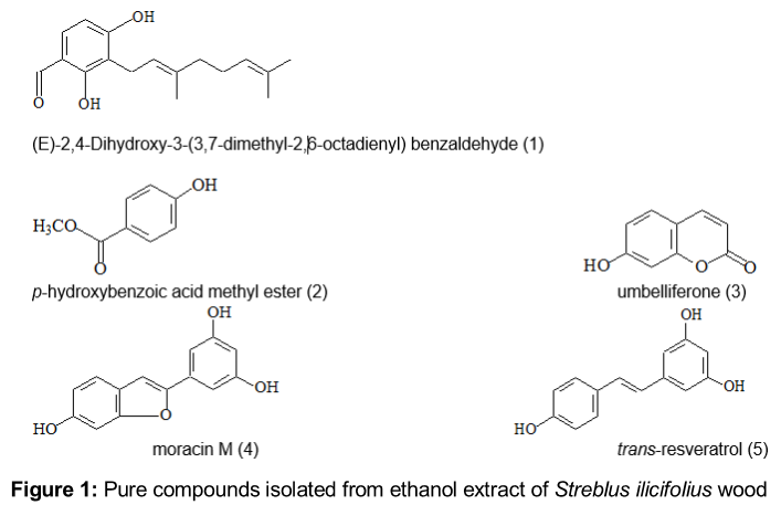
Five pure compounds were isolated from ethanol extract of S. ilicifolius (). The structures of the isolated compounds, identified by physical properties and spectroscopic data, including UV, IR, NMR and MS data and confirmed by comparison with previously reports are as (E)-2,4-dihydroxy-3-(3,7-dimethyl-2,6-octadienyl) benzaldehyde (1) [14], p-hydroxy benzoic acid methyl ester (2) [15], umbelliferone (3) [16], moracin M (4) [17] and trans-resveratrol (5) [18]. Their spectral data are given as follow:
(E)-2,4-Dihydroxy-3-(3,7-dimethyl-2,6-octadienyl) benzaldehyde (Compound 1); C17H22O3
Compound 1 was obtained as colorless needles, soluble in chloroform. The melting point showed at 72.33 ºC (Differential scanning calorimeter). The EI mass spectrum of compound 1 exhibited a molecular ion at 274 m/z, consistent with a molecular formula of C17H22O3, confirmed by HR-EIMS which showed a molecular ion peak at m/z 274.1560 calcd. for C17H22O3, 274.1566. The UV spectrum in chloroform demonstrated absorption maximum in chloroform at λmax (absorbance) 228 (0.180), 242 (0.235), 286(0.593) and 396(0.007) nm. The IR spectrum exhibited maximum absorption bands at 3401, 2923, 2851 and 1622 cm-1. The 1H and 13C NMR spectra of compound 1 are showed as:
1H NMR (500 MHz, CDCl3): 1.57 (3H, s, H-9´), 1.65 (3H, s, H-10´), 1.80 (3H, s, H-4´), 2.06 (4H, m, H-5´, H-6´), 3.43 (2H, d, J=7.3 Hz, H-1´), 5.02 (1H, m, H-7´), 5.24 (1H, tq, J=7.3, 1.1 Hz, H-2´), 6.28 (1H, s, 4-OH), 6.46 (1H, d, J=8.4 Hz, H-5), 7.28 (1H, d, J=8.4 Hz, H-6), 9.66 (1H, s, 1-COH), 11.75 (1H, s, 2-OH);
13C NMR (125 MHz, CDCl3): 16.23 (C-16´), 17.69 (C-9´), 21.33 (C-1´), 25.65 (C-10´), 26.26 (C-6´), 39.67 (C-5´), 109.04 (C-5), 113.50 (C-3), 115.02 (C-1), 120.55 (C-2´), 123.60 (C-7´), 132.16 (C-8´), 133.50 (C-6), 140.06 (C-3´), 161.65 (C-4), 162.70 (C-2), 194.63 (1-COH).
p-Hydroxybenzoic acid methyl ester (Compound 2); C8H8O3
The compound 2 was obtained as a colorless needles, soluble in methanol and dimethyl sulfoxide. The melting point showed at 118 °C. The % abundance GC-MS of compound 2 exhibited a molecular ion at 152 m/z, consistent with a molecular formula of C9H6O3. The UV spectrum in methanol demonstrated absorption maximum in methanol at λmax (absorbance) 255 (0.837) and 314 (0.121) nm. The IR spectrum exhibited maximum absorption bands at 3355, 1681, 1609, 1585 and 1513 cm-1. The 1H and 13C NMR spectra of compound 2 are showed as:
1H-NMR (300 MHz, DMSO-d6): 3.78 (3H, s, 7-OCH3), 6.84 (2H, d, J=8.70 Hz, H-2, H-6), 7.80 (2H, d, J=8.68 Hz H-3, H-5,), 10.34 (1H, brs, 4-OH);
13C-NMR (75 MHz, DMSO-d6): 52.06 (7-OCH3), 115.78 (C-2, C-6), 120.68 (C-1), 131.86 (C-3, C-5), 162.39 (C-4), 166.50 (C-7).
Umbelliferone (Compound 3); C9H6O3
The compound 3 was obtained as a yellowish crystalline, soluble in methanol and dimethyl sulfoxide. The melting point showed at 230 °C. It gave a molecular ion at 162 m/z in the % abundance GC-MS, suggesting a tentative molecular formula of C9H6O3. The UV spectrum in methanol displayed absorptions maximum in methanol at λmax 216, 258 and 332 nm. The 1H and 13C NMR spectra of compound 3 are showed as:
1H-NMR(300 MHz, DMSO-d6): 6.19 (1H, d, J = 9.45 Hz, H-3), 6.71 (1H, d, J=2.28Hz, H-8), 6.78 (1H, dd, J=8.49, 2.31 Hz, H-6), 7.52 (1H, d, J=8.49 Hz, H-5), 7.93 (1H, d, J=9.48 Hz, H-4);
13C-NMR(75 MHz, DMSO-d6): 102.61 (C-8), 111.66 (C-4a), 111.77 (C-3), 113.62 (C-6), 130.16 (C-5), 145.01 (C-4), 155.97 (C-8a), 160.91 (C-7), 161.89 (C-2).
Moracin M (Compound 4); C14H10O4
The compound 4 was obtained as a cream needles, soluble in methanol and dimethyl sulfoxide. The melting point showed at 270 °C. It gave a molecular ion at 242 m/z in the EI mass spectrum, corresponding to C14H10O4. The UV spectrum in methanol showed absorptions at λmax (absorbance) 216 (0.863), 315 (0.938) and 328 (0.796) nm. The IR spectrum showed the absorption bands at 3521, 3328, 1613, 1436, 1292 and 1141 cm-1. The 1H and 13C NMR spectra of compound 4 are showed as:
1H-NMR (500 MHz, DMSO-d6): 6.20 (1H, t, J=2.05 Hz, H-4´), 6.67 (2H, d, J=2.05Hz , H-2´, H-6´), 6.72 (1H, dd, J=8.46, 2.06 Hz, H-5), 6.91 (1H, ddd, J=2.06, 0.91 Hz, H-7), 7.05 (1H, d, J=0.91 Hz, H-3), 7.37 1H, d, J=8.25 Hz, H-4), 9.39 (2H, brs, 3´-OH, 5´-OH), 9.95 (1H, brs, 6-OH);
13C-NMR (125 MHz, DMSO-d6): 97.62 (C-7), 101.69 (C-3), 102.47 (C-2´, C-6´), 102.81 (C-4´), 112.61 (C-5), 120.94 (C-3a), 121.26 (C-4), 131.82 (C-1´), 154.13 (C-2), 155.42 (C-7a), 155.88 (C-6), 158.94 (C-3´, C-5´).
trans-Resveratrol (Compound 5); C14H12O3
The compound 5 was obtained as a brownish crystalline, soluble in methanol and dimethyl sulfoxide. The melting point showed at 265 °C. It gave a molecular ion at 228 m/z in the % abundance GC-MS, suggesting a tentative molecular formula of C14H12O3. The UV spectrum in methanol, showed absorptions at λmax (absorbance) 207 (0.647), 315 (0.531) and 328 (0.450) nm. The IR spectrum exhibited maximum absorption bands at 3234, 1618, 1578, 1508 and 1485 cm-1. The 1H and 13C NMR spectra of compound 5 are showed as:
1H-NMR(300 MHz, DMSO-d6): 6.10 (1H, brs, J=2.10 Hz, H-4), 6.37 (2H, d, J=2.13 Hz, H-2, H-6), 6.74 (2H, d, J=8.58 Hz, H-3´, H-5´), 6.80 (1H, d, J = 16.22 Hz, H-α), 6.93 (1H, d, J= 16.35 Hz, H- α´), 7.38 (2H, d, J=8.55 Hz, H-2´, H-6´), 9.20 (2H, brs, 3-OH, 5-OH), 9.56 (1H, brs, 4´-OH);
13C-NMR (75 MHz, DMSO-d6): 102.19 (C-4), 104.72 (C-2, C-6), 115.94 (C-3´, C-5´), 126.07 (C-α), 128.29 (C-2´, C-6´, C-α´), 128.49 (C-1´), 139.69 (C-1), 157.65 (C-4´), 158.93 (C-3, C-5).
GC-MS (EI): m/z (%) = 228.1[M+] (100), 211.1 (10), 181.1 (17), 157.1 (12), 115.0 (9), 44.0(12).
Antityrosinase activity of isolated compounds
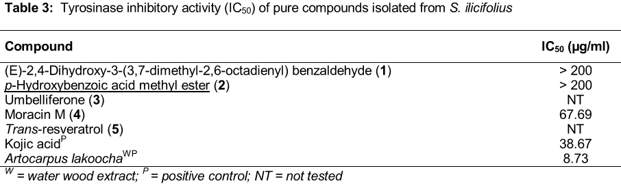
Determination of tyrosinase inhibitory activity by dopachrome method of isolated compounds was showed in .
Antimicrobial activity of isolated compounds
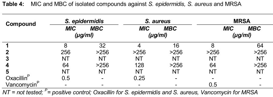
From the screening of antimicrobial activity, S. ilicifolius ethanol extract displayed an inhibition zone against S. aureus and S. epidermidis. MIC and MBC of isolated compounds were determined with S. epidermidis, S. aureus and methicillin-resistant S. aureus (MRSA). The MIC and MBC values of isolated compounds are showed in .
Discussion
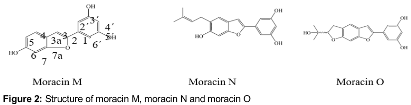
Five pure compounds were isolated from S. ilicifolius for the first time although all of them are known compounds. Compound 1 and 2 were inactive with antityrosinase activity. While, compound 4 was found to exhibit a high antityrosinase activity. The hydroxyl group at the 6 position of moracin M has been reported that it might mediate the inhibitory activity [19]. Moracin M is 2-arylbenzofuran, the hydroxyl group at the 6 position might mediate the inhibitory activity, compared with two 2-arylbenzofurans; moracin N and moracin O (). Moracin N and moracin O belong to isoprenyl-substituted 2-arylbenzofuran which showed the activity against tyrosinase with IC50 30.52 µM and 93.58 µM, respectively. In moracin O, the isoprenyl group forms a five-membered ring with the hydroxyl group at the 6 position, whereas in moracin N, the isoprenyl group remains intact which might contribute to its higher tyrosinase inhibitory activity than moracin O [19].
The activity of tyrosinase inhibitors are classified into four types, including competitive inhibitors, uncompetitive inhibitors, mixed type (competitive/ uncompetitive) inhibitors, and non-competitive inhibitors [26]. Resveratrol is a polyphenolic phytoalexin that belongs to the stilbenes, which have demonstrated potent tyrosinase inhibitory activity [27]. This substance acts as a “suicide substrate” for tyrosinase [28]. While, umbelliferone is a coumarin analog with hydroxyl group at C7 could be competitive potent tyrosinase inhibitory activity like the other coumarin, esculetin [29]. Moreover moracin M, the arylbenzofuran showed competitive potent tyrosinase inhibitory activity [30].
(E)-2,4-Dihydroxy-3-(3,7-dimethyl-2,6-octadienyl) benzaldehyde (1) showed strong inhibitory effect against S. epidermidis, S. aureus and MRSA. Friedman et al [25]. reported that 2, 3-dihydroxybenzaldehyde was highly active against four bacteria (Campylobacter jejuni, Escherichia coli, Listeria monocytogenes and Salmonella enterica) because hydroxyl group at the position 2 and 3 enhance the antimicrobial activities of benzaldehyde. Then, hydroxyl group at the position 2 and 3 of 2, 3- dihydroxy-4-geranyl benzaldehyde might mediate the inhibitory activity.
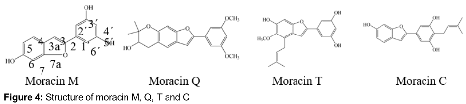
Moracin M (4) is the arylbenzofuran, showed weak inhibitory effect against, S. epidermidis, S. aureus and MRSA. Mazimba et al [30] reported moracin M against S. aureus (MIC 62.5 µg/mL), B. subtilis (MIC 31.25 µg/mL), M. flavus (MIC 125 µg/mL), S. faecalis (MIC 62.5 µg/mL), S. abony (MIC 62.5 µg/mL) and P. aeruginosa (MIC 125 µg/mL). Moreover, Kuetu et al [32] reported that all tested microbial species (Gram-positive, Gram-negative bacteria and fungi) were inhibited by non-prenylated arylbenzofurans; moracin M and moracin Q () weaker than prenylated arylbenzofurans; moracin T and moracin C () because prenyl increases the antimicrobial activity of arylbenzofurans. p-hydroxybenzoic acid methyl ester (2) showed very low MIC against S. epidermidis. It is one of a homologous series of parabens used to exert antimicrobial affect especially, useful against molds and yeasts. It againsts A. oryzae (MIC 600 µg/mL), T. lignorum (MIC 250 µg/mL), S. lutea (MIC 4000 µg/mL), E. cloacae (MIC 1000 µg/mL), P. vulgaris (MIC 2000 µg/mL) (Kibbe, 2000), E. coli (wild type) (MIC 1400 µg/mL), E. coli (envelope mutant) (MIC 1000 µg/mL), P. aeruginosa (wild-type) (MIC 1800 µg/mL) and P. aeruginosa (envelope mutant) (MIC 1000 µg/mL) [33].
Some of the isolated compounds; umbelliferone (3) and trans-resveratrol (5) did not test because they have trace amount. However, they have been reported of anti-microbial activity.
Resveratrol showed activity against Gram-positive bacteria higher than Gram- negative bacteria. It had the highest activity against Bacillus cereus followed by Staphylococcus aureus, MRSA and Enterococcus faecalis [34]. Morever, Kukric and Topalic-Trivunovic [35] reported that trans-resveratrol showed antimicrobial activity against test organisms (E. coli, B. subtilis, Straphylococcus sp. and S. lutea) higher than cis-resveratrol. In addition, trans-resveratrol displayed potent antifungal activity against human pathogenic fungi (C. albicans, S. cerevisiae and T. beigelii with MIC 10, 10-20 and 10 µg/mL), respectively [36].
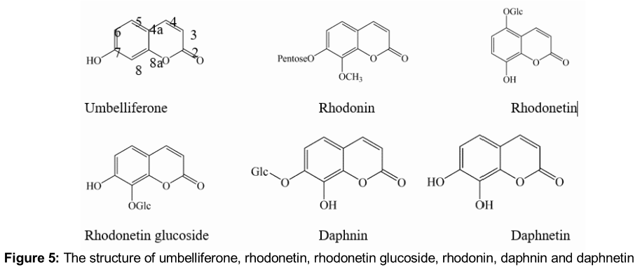
Umbelliferone (), which has a free hydroxyl at the position 7, did not possess good antibacterial activity while, daphnetin which has free hydroxyl at the position 7 and 8, is the most active compound out of all the coumarins tested. Moreover, daphnin and rhodonetin which has free hydroxyl at the position 8, showed better activity than rhodonetin glucoside which the glycoside at the position 8 and rhodonin which methoxyl at the position 8. From the data concluded that coumarins with free hydroxyl at the position 8 possess better antibacterial activity [37].
However, these results will be useful for further study for pharmaceutical cosmetics or application for skin treatment of hyperpigmentation and infectious diseases such as the formulations of cream, serum, mask and so on.
Conclusion
Five pure compounds were isolated from the wood of S. ilicifolius. This is the first report on the biological activities and phytochemical profile of S. ilicifolius. Also, (E)-2,4-Dihydroxy-3-(3,7-dimethyl-2,6-octadienyl) benzaldehyde from natural products, which has been investigated here for the first time, shows good antimicrobial activity. Moracin M has antityrosinase activity. However, further studies ascertain the actual therapeutic potentials of the compounds in the management of infectious diseases and hyperpigmentation.
Declarations
Acknowledgement
References
Archives


News Updates